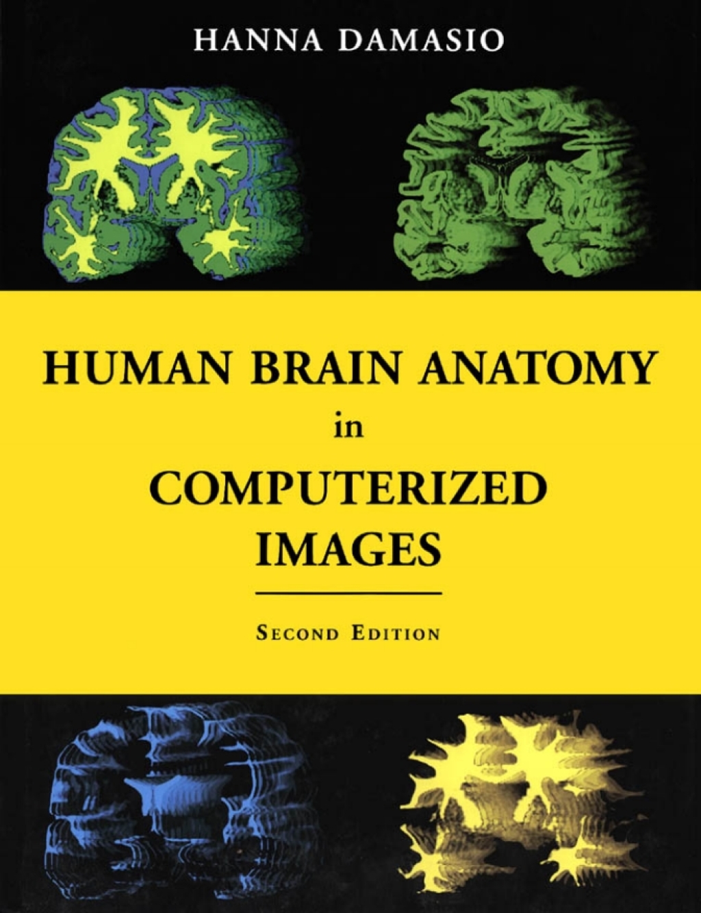
By using non-invasive tomographic scans, modern neuroimaging technologies are revealing the structure of the human brain in unprecedented detail. This spectacular progress, however, poses a critical problem for neuroscientists and for practitioners of brain-related professions: how to find their way in the current tomographic images so as to identify a particular brain site, be it normal or damaged by disease? Prepared by a leading expert in advanced brain-imaging techniques, this unique atlas is a guide to the localization of brain structures that illustrates the wide range of neuroanatomical variation. It is based on the analysis of 29 normal human brains obtained from three-dimensional reconstructions of magnetic resonance scans of living persons. The Second Edition of this atlas offers entirely new images, all from new brain specimens.
“Diagnostic Imaging: Oral and Maxillofacial 3rd Edition” has been added to your cart. View cart
Human Brain Anatomy in Computerized Images 2nd Edition
Author(s): Hanna Damasio M.D.
Publisher: Oxford University Press
ISBN: 9780195165616
Edition: 2nd Edition
$39,99
Delivery: This can be downloaded Immediately after purchasing.
Version: Only PDF Version.
Compatible Devices: Can be read on any device (Kindle, NOOK, Android/IOS devices, Windows, MAC)
Quality: High Quality. No missing contents. Printable
Recommended Software: Check here

