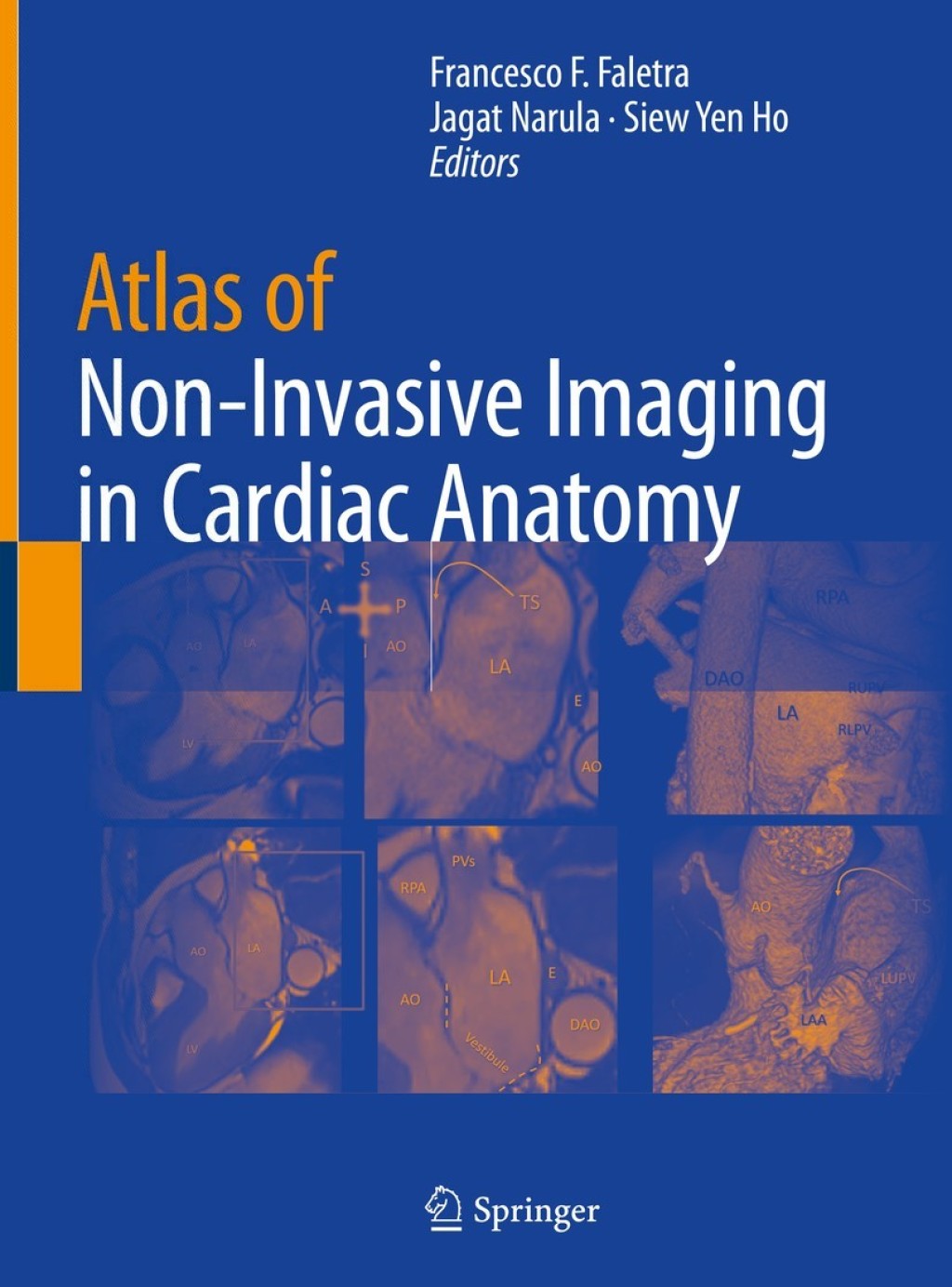
This atlas provides a detailed visual resource of how sophisticated non-invasive imaging relates to the anatomy observed in a variety of cardiovascular pathologies. It includes investigation of a wide range of defects in numerous cardiac structures. Mitral valve commissures, atrioventricular septal junction and right ventricular outflow tract plus a wealth of other structures are covered, offering readers a comprehensive integrative experience to understand how anatomic subtleties are revealed by modern imaging modalities. Atlas of Non-Invasive Imaging in Cardiac Anatomy provides a detailed set of visual instructions that is of use to any cardiovascular professional needing to understand the orientation of a patient’s imaging. Therefore this is an essential guide for all trainee and practicing cardiologists, cardiac imagers, cardiac surgeons and interventionists.
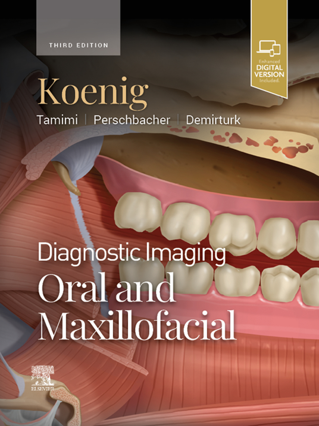
“Diagnostic Imaging: Oral and Maxillofacial 3rd Edition” has been added to your cart. View cart
Atlas of Non-Invasive Imaging in Cardiac Anatomy 1st Edition
Author(s): Francesco F. Faletra; ‎Jagat Narula; ‎Siew Yen Ho
Publisher: Springer
ISBN: 9783030355050
Edition: 1st Edition
$39,99
Delivery: This can be downloaded Immediately after purchasing.
Version: Only PDF Version.
Compatible Devices: Can be read on any device (Kindle, NOOK, Android/IOS devices, Windows, MAC)
Quality: High Quality. No missing contents. Printable
Recommended Software: Check here

