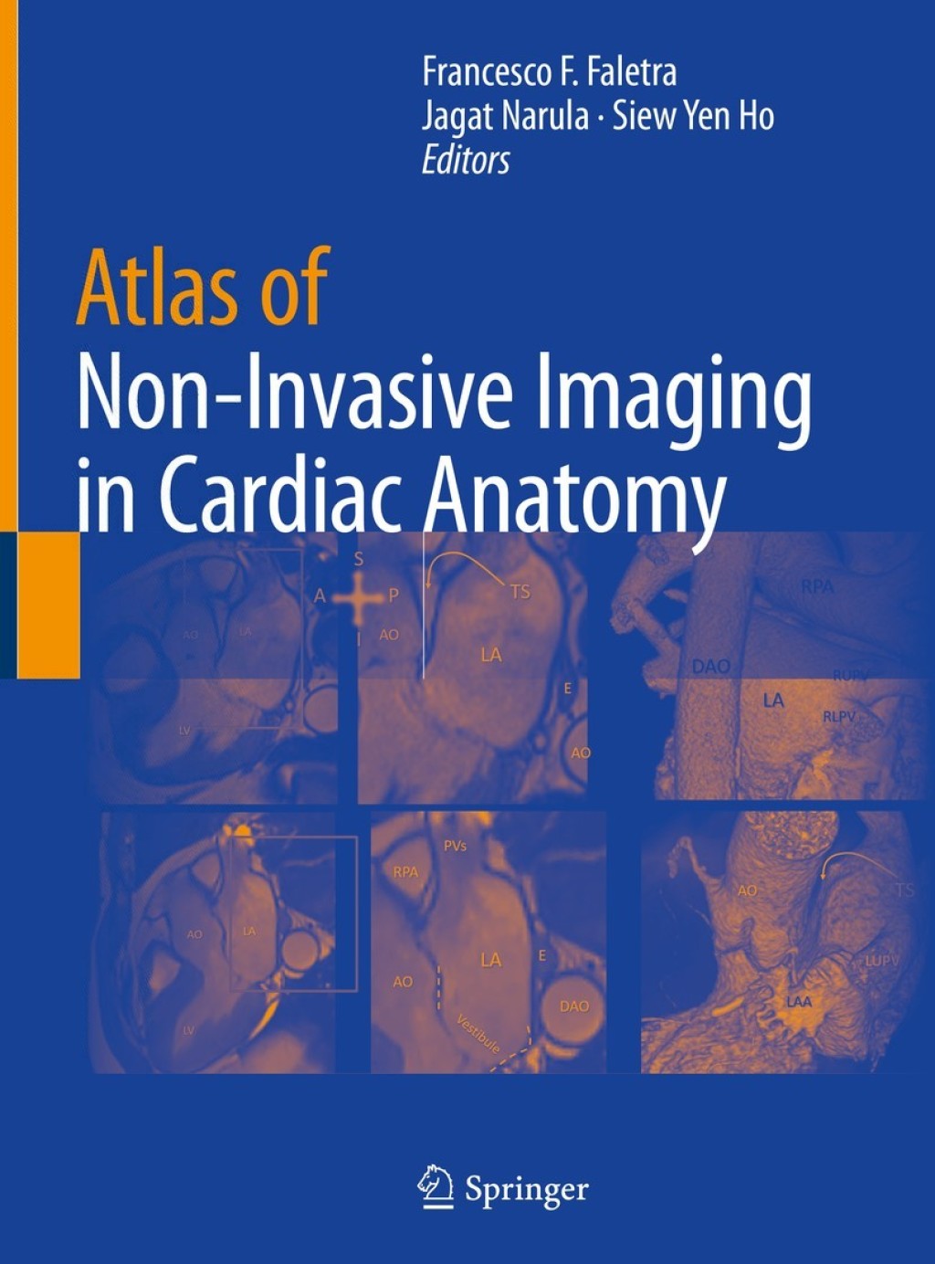
This atlas provides a detailed visual resource of how sophisticated non-invasive imaging relates to the anatomy observed in a variety of cardiovascular pathologies. It includes investigation of a wide range of defects in numerous cardiac structures. Mitral valve commissures, atrioventricular septal junction and right ventricular outflow tract plus a wealth of other structures are covered, offering readers a comprehensive integrative experience to understand how anatomic subtleties are revealed by modern imaging modalities. Atlas of Non-Invasive Imaging in Cardiac Anatomy provides a detailed set of visual instructions that is of use to any cardiovascular professional needing to understand the orientation of a patient’s imaging. Therefore this is an essential guide for all trainee and practicing cardiologists, cardiac imagers, cardiac surgeons and interventionists.
“Genetics and Neurology” has been added to your cart. View cart
Atlas of Non-Invasive Imaging in Cardiac Anatomy 1st Edition
Author(s): Francesco F. Faletra; ‎Jagat Narula; ‎Siew Yen Ho
Publisher: Springer
ISBN: 9783030355050
Edition: 1st Edition
$39,99
Delivery: This can be downloaded Immediately after purchasing.
Version: Only PDF Version.
Compatible Devices: Can be read on any device (Kindle, NOOK, Android/IOS devices, Windows, MAC)
Quality: High Quality. No missing contents. Printable
Recommended Software: Check here

